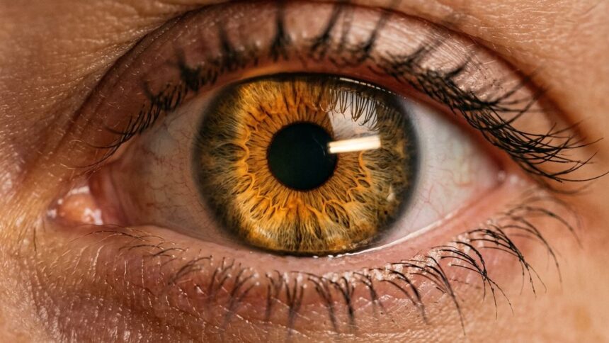
Most people know the primary optical rule of the iris: brighter illumination constricts the pupil, while dim light expands it. Yet optical lighting explains only a fraction of pupillary dynamics across an ordinary day. In cognitive neuroscience and psychophysiology, the pupil functions as a sensitive real-time window into the autonomic nervous system—subtly dilating in response to cognitive workload, sensory salience, and physiological arousal. The textbook rule explains a fraction of what the aperture does: across an ordinary evening your pupil varies by a factor of two or more, and only part of that travel is bought with photons. The rest is modulated by autonomic arousal pathways — driven by ambient illumination, cognitive effort, and sympathetic tone — producing subtle shifts that reflect physiological arousal rather than a single specific emotion.
Sixty years of pupillometry have traced why pupils dilate under emotional load, and the trace forms an anatomy — a five-node autonomic circuit running from a thin column of blue-gray neurons at the brainstem’s floor, through the midbrain nucleus owning the pupil’s brake, into the two antagonistic iris muscles, and out into the space between two faces — where pupils subtly reflect mutual engagement and interpersonal attunement through autonomic micro-adjustments. This article follows that circuit node by node — the full specification of why pupils dilate.
Beyond the Light Reflex: How Emotional Salience Hijacks the Iris
The pupil’s day job is photometry, and the mechanism is one of neurology’s cleanest. Melanopsin-bearing retinal ganglion cells tally the photon budget and cable the sum to the pretectal olivary nucleus; the signal crosses to the paired Edinger-Westphal nuclei, rides the oculomotor nerve to the ciliary ganglion, and lands on the sphincter pupillae, the ring of muscle that squeezes the aperture shut. Constriction starts roughly 200 milliseconds after a light step, so reliable that pupil size in controlled light remains one of the few signs clinicians trust in sedated patients — the floor, but only the floor, of why pupils dilate.
Emotional salience crosses that mechanism like express traffic on a country lane — the first detour in why pupils dilate. Bradley, Miccoli, Escrig and Lang’s laboratory showed it cleanly: affectively charged pictures widened observers’ pupils while screen luminance was held constant [Bradley et al., 2008]. The change scaled with rated arousal, not valence: a threat and a couple embracing opened the aperture comparably, both crossing the brain’s threshold of this matters now. That threshold, not the light meter, decides why pupils dilate when one person’s gaze settles on another’s face.
The contest between light and affective drive is adjudicated, not random: at equal photic input salience wins through release of the parasympathetic brake, upstream of the Edinger-Westphal pipeline whose cross-talk into neighboring autonomic channels is documented in the photic sneeze reflex. A bright sky and a noticed face pull on the same muscarinic line in opposite directions, and under load the face wins — which is why pupils dilate for people in rooms whose lighting has not changed at all.
Node 2: The Edinger-Westphal Override — Releasing the Parasympathetic Brake
Dissected, the override is disinhibition: Edinger-Westphal neurons answer to more than photons, sitting downstream of limbic drives that suppress the cholinergic outflow as arousal climbs. The brake line goes slack, the sphincter relaxes, and the pupil widens on the very light budget it was rationing. Loewenfeld’s The Pupil reads this two-sided control as ancient vertebrate equipment, and modern reviews confirm it [Mathôt, 2018]: why pupils dilate for interesting faces while constricting briskly to a flashlight a few inches away is a released brake, not a changed photic budget.
The Locus Coeruleus-Norepinephrine Axis: The Chemical Trigger of Attraction
Salience needs a transmitter, and the transmitter of record is norepinephrine, broadcast from the locus coeruleus — a bar of deeply pigmented neurons on the floor of the fourth ventricle holding near-monopoly on central norepinephrine. Its axons vend the chemical to virtually every cortical sheet, diffusely enough that much of its transmission is volumetric — signal spreading past the synapse to receptor fields. When the LC fires, the forebrain’s gain moves; when it idles, the gain drops. One nucleus, one transmitter, everywhere at once — its verdict on any salient face is the second half of why pupils dilate.
The authoritative account of what that architecture is for is Aston-Jones and Cohen’s adaptive gain theory [Aston-Jones & Cohen, 2005]. The LC runs in two modes that together explain why pupils dilate in opposite registers. Tonic mode — slow, irregular firing — is exploration: attention spreads, distraction tolerated. Phasic mode — brief, high-amplitude bursts driven by task and reward evaluation — is engagement: cortical responsiveness rises, discrimination sharpens, and behavior locks onto a target.

The theory’s economics are blunt: the LC computes whether the world warrants engagement and stamps the verdict on a pupil whose diameter under constant light tracks its firing mode. At a first meeting the sequence reads like a specification — a face crosses the salience threshold, dopaminergic reward signals of the kind that make music give you goosebumps tip the LC phasic, norepinephrine floods the cortical bed, the eye widens. In miniature, that chain is why pupils dilate within the first two seconds of noticing someone attractive.
Norepinephrine is not a love drug; it is a gain drug, and the same phasic burst serves attraction, a rising siren, a jackpot, a crisis — the LC invoices every salient event at the same rate. What makes attraction legible rather than interchangeable with alarm is limbic context — facial processing, reward prediction, memory — around one identical pulse. The pupil broadcasts the invoice’s size, never its itemization.
Node 1: The LC-NE Gain Switch — A Salience Amplifier Behind the Eyes
Read as circuitry, the LC-NE axis is an amplifier between perception and behavior with one public meter wired to it: under fixed illumination the pupil reads that meter — phasic mode wide, tonic rest narrow — trustworthy enough that neuroscientists treat pupil diameter as a dependent variable. This is the first and central node of why pupils dilate for attractive faces: the aperture is where invisible appraisal economics becomes a public event.
Autonomic Pupillary State: Basal Rest vs Affective Salience Activation
- Core Focus: Mechanism: The retinal photon budget sustains pretectal drive; the constrictor ring keeps its resting squeeze with no descending interference.
- Neurochemistry: Autonomic State: The locus coeruleus idles in slow tonic mode; sympathetic outflow holds near floor; global cortical gain sits at baseline.
- Dynamic: Subjective State: Attention is diffuse and inward; peripheral events fail to claim detection priority; the pupil reads small and stable.
Biomechanical Tug-of-War: Dilator Pupillae vs. Iris Sphincter
The iris is the only tissue where two smooth muscles arm-wrestle continuously over the same square millimeter, and the contest is architectural. Ringing the pupil’s edge runs the sphincter pupillae, a thin annulus whose contractile life is spent tightening the aperture. Spreading outward to the iris root lies the dilator pupillae, a radial myoepithelial sheet whose contraction pulls the pupillary margin outward, opening the eye. The two are mechanically opposed, and under healthy dual innervation the aperture lands wherever the moment’s balance dictates — the biomechanical floor of why pupils dilate at all.
The controllers arrive from different floors of the neuraxis. The sphincter answers to the pipeline already traced: Edinger-Westphal outflow, muscarinic drive at the margin. The dilator answers to a sympathetic arc three relays longer — hypothalamus to the ciliospinal center at C8–T2, the superior cervical ganglion, then through the long ciliary nerves to alpha-1 receptors on the radial sheet. Under emotional salience both strings move at once — the dilator pulled, the parasympathetic brake released — which is why pupils dilate smoothly and in graded steps rather than in a snap.
Clinical anatomy demonstrates both autonomic pathways are active. In Horner’s syndrome an interrupted chain leaves one pupil small and unable to dilate fully — the dilator is a wired, independent effector, not slack behind a relaxing sphincter. The muscles’ physics also differ: ring squeeze is short-path and fast; the radial draw crosses the full iris and takes visibly longer. That asymmetry is part of why pupils dilate under affect on a curve readable from across a dinner table — measurement, not metaphor.
Node 3: The Iris Duel — Radial Draw Against Ring Squeeze
As a circuit element, this node is where chemistry becomes geometry: commands converge on one pigmented membrane from opposite autonomic towers, and the aperture you can read without instruments is the running arithmetic of their collision. Several hundred times an evening, why pupils dilate is an argument two muscles settle in fractions of a second.
Pupillary Contagion: The Unconscious Mirroring That Builds Attraction
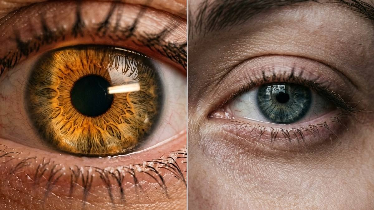
Attraction is the rare reflex with a social exponent: your pupil does not merely respond to a face — it responds to another pupil’s response. Kret, Fischer and De Dreu documented the mechanism under laboratory control [Kret et al., 2015]: participants watched videos of faces whose pupils visibly dilated or constricted, and the observers’ own pupils synchronized with the change, mimicry emerging within roughly a second. The effect was selective — strong for in-group partners, reduced for out-group faces — and transactional: participants whose pupils had mimicked an in-group partner’s dilation later trusted that partner more. Pupil mimicry predicted behavior, and it is the social branch of why pupils dilate.
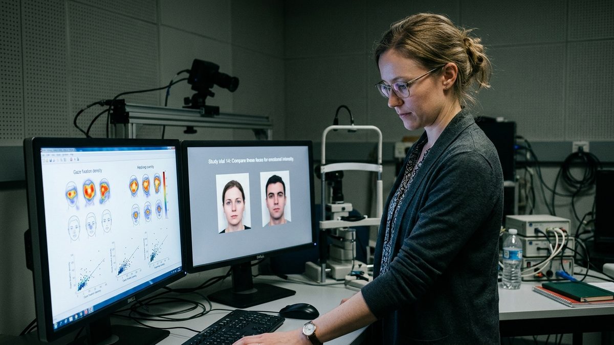
The reading is autonomic empathy: why pupils dilate is as much the observer’s business as the observed person’s. Pupillary contagion belongs to the family that contains contagious yawning and facial mimicry — another person’s internal state routed into one’s own autonomic apparatus without instructions. The candidate circuitry fits the route: visual cortex and frontal eye fields extract the partner pupil’s geometry; the insula, which interocepts one’s own autonomic state, plausibly maps it back onto the self; and oxytocin, implicated across gaze and trust paradigms, is a proposed facilitator of the attunement — a hypothesis, not yet a mechanism.
Measured, not proposed, is the outcome: a widening pupil is information about interest and warmth, absorbed without awareness. It is also where interpretation gets dangerous — the misattribution documented in the suspension bridge effect psychology waits inside every widened pupil, fear-born physiology misfiling itself as romance.
Why pupils dilate in the eyes of someone whose heart just raced may owe nothing to romance and everything to adrenaline — a coaster, cold air, strong coffee. The pupil transmits arousal’s amplitude with high fidelity and no semantic label. It is a broadcast, not a confession.
Node 4: The Mimicry Engine — Social Attunement and Pupillary Contagion
As circuitry, mimicry is the loop that closes between two nervous systems: visual extraction, insular mapping, a copycat adjustment — then it runs again both ways until two apertures settle into mutual register. This is where why pupils dilate acquires its social meaning: the first autonomic fact two people exchange while still deciding whether they like each other.
From Hess to Infrared Pupillometry: 60 Years of Measuring Why Pupils Dilate
The field that measures why pupils dilate was founded in 1960 by a Gestalt psychologist with a darkroom. Eckhard Hess at the University of Chicago suspected the pupil exposed interest directly, and with student Polt he tested it with telecentric photographs under fixed illumination, mental multiplication, and emotionally graded pictures [Hess & Polt, 1960]. Dilation tracked both load and interest — harder arithmetic enlarged the pupil further — and later demonstrations pushed the finding to its social edge: a woman’s photograph retouched to enlarge her pupils drew more favorable judgments than its small-pupil twin, and observers could not say why. The pupil had told them; nobody had.
Hess’s instrumentation aged badly — variability was large, samples small, light easy to confound — and the field spent decades in quarantine. What arrived from the 1980s onward, maturing in the 2010s, was infrared video-oculography: eye trackers imaging the pupil in light the eye cannot see, sampling its diameter thirty to a thousand times a second while logging luminance, so photic influence can be partitioned out of every change [Mathôt, 2018]. The signal is reliable; the interpretation is the hard part — which is why every study since Hess begins by asking why pupils dilate.
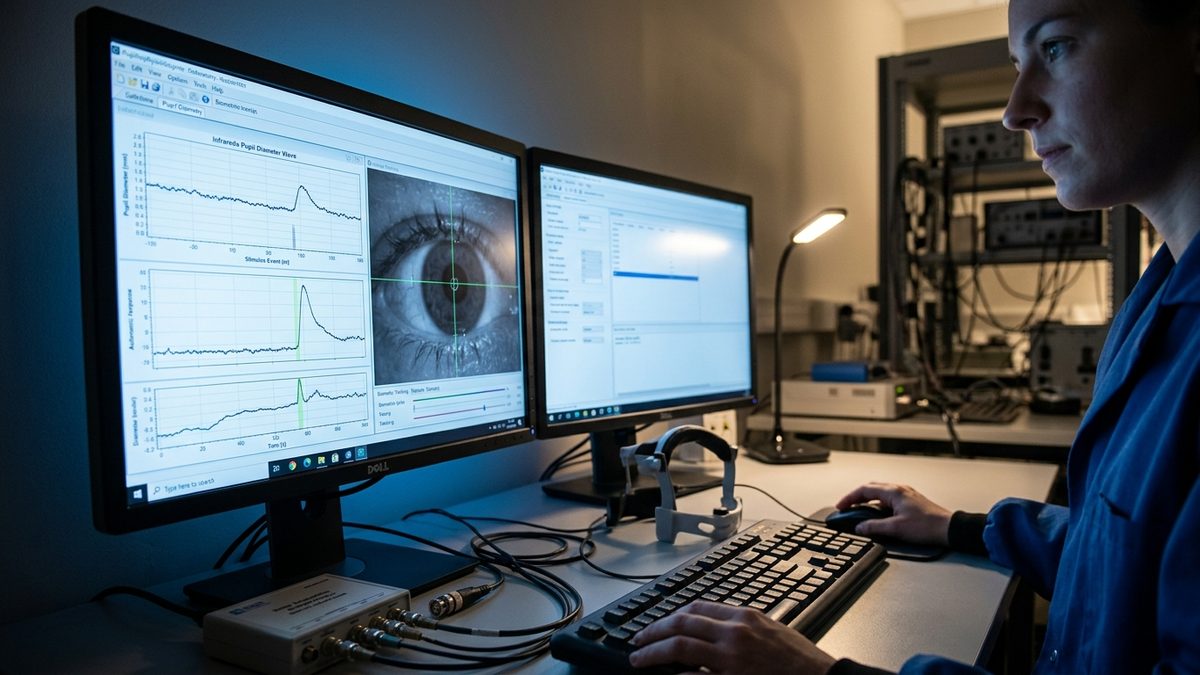
What the modern apparatus exposed most sharply is the split. Cognitive effort produces dilation strikingly alike in kind — the multiplication result has repeated under every infrared generation — but its physiognomy differs: task-evoked dilation arrives locked to a stimulus, peaks within seconds, decays with the trial, and rides the phasic LC mode. Affective dilation accumulates with sustained emotional load and co-varies with other sympathetic outlets — heart rate, skin conductance — the involuntary family tracked in the body’s signals of fatigue before collapse. Time course and companionship separate the two; honesty does not. The honest answer to why pupils dilate has two heads — effort and salience — and the pupil confesses both with equal credibility.
Node 5: Load vs. Salience — What the Pupil Confesses, and What It Cannot
The pupil never lies in the precise sense that it reports arousal without editorial approval; where it disappoints romantics is that honesty is not specificity. A millimeter of widening is as true of arithmetic as of attraction, which is every forensic claim’s undoing: the pupil proves desire dies against the record. The disciplined question is not whether the aperture moved but when, in company with which signals, at what luminance — the three coordinates on which why pupils dilate becomes readable instead of romantic. Asked that way, the aperture is an instrument — the same instrument two nervous systems already run on each other, from across the table.
“Pupils only dilate during conscious sexual desire or romantic attraction — a widening pupil certifies a crush with complete fidelity.”
Affective pictures of fear, thrill or disaster dilate pupils with no romance involved (Bradley et al., 2008); effortful mental arithmetic dilates them too (Hess and Polt, 1960); bright light constricts them at identical luminance. The pupil reports salience and autonomic arousal — never the caption the observer wishes to assign.
Autonomic Pupillary Response & Neuro-Ophthalmic Modality Matrix
The matrix below sets four pupil states across the five neuro-ophthalmic parameters separating them. Attraction and effort share a dilation route but differ in timing; the light reflex and shock talk precision in opposite directions — an opposition that explains why pupils dilate four different ways.
| Physiological State | Affective Attraction (Mydriasis) | Cognitive Processing Load | Pupillary Light Reflex (Miosis) | Pathological / Sympathetic Shock |
|---|---|---|---|---|
| Primary Neural Pathway | Limbic amygdala-hypothalamic input driving locus coeruleus bursts and downstream sympathetic outflow | Prefrontal-parietal task circuitry engaging phasic LC-NE firing locked to stimulus events | Melanopsin retinal ganglion cells to pretectal olivary nucleus to Edinger-Westphal via oculomotor nerve | Adrenal medullary catecholamine surge with global sympathetic activation and loss of local grading |
| Biomechanical Muscle Target | Dilator pupillae alpha-1 contraction plus simultaneous sphincter relaxation via E-W disinhibition | Dilator-dominated slow shift with a minor sphincter tone decrement | Sphincter pupillae M3 muscarinic contraction, bilateral, with high temporal precision | Dilator mass activation by circulating epinephrine; course irregular and often asymmetric |
| Autonomic Tone Dominance | Sympathetic gain with the parasympathetic brake released simultaneously — dual-string sweep | Phasic sympathetic engagement over a normal parasympathetic baseline | Parasympathetic tone dominance; sympathetic outflow held near floor | Overwhelming sympathetic dominance; parasympathetic counter-pull negligible |
| Mean Pupil Diameter Delta (mm) | +0.4 to +1.5 typical at matched luminance, waxing with sustained engagement | +0.3 to +1.0, event-locked, decaying to baseline within seconds after the trial | −2 to −5: from roughly 5-6 mm ambient down to 2-3 mm in ordinary room light | +2 to +4: apertures commonly 6-8 mm in catecholamine surge or shock states |
| Subconscious Social Signal | Interest, warmth, availability — read within a second and mimicked by the observer | Commitment and engagement — often read as attentive, not necessarily as favor | Neutral to the interaction; occasionally misread as coolness at close range | Alarm broadcast: fixed mid-position pupils alongside pallor accelerate helping behavior |
Any single reading of why pupils dilate is incomplete by construction. Attraction and arithmetic both buy dilation at matched luminance; the light reflex solves for the opposite sign, and shock broadcasts in catecholamines. The honest reading keeps all four columns in view.
The Neurological Signal: What Pupil Dilation Actually Communicates
Why pupils dilate when you look at someone attractive is, on the record, a five-node event: a blue nucleus votes on engagement, norepinephrine raises the brain’s gain, the parasympathetic brake releases, the radial muscle draws while the ring eases — and across the table your partner’s pupil copies the widening, social attunement shifting accordingly. Light sets the floor of the reading, salience moves the dial, and nobody moves the dial by wanting to.
Read without romance’s shadow, the rule holds: widened pupils under constant light mean real arousal and interest in engaging — but interest is not a census of categories, and the same widening rides adrenaline, arithmetic, listening, and attraction alike. Amplitude and latency are governed by subcortical autonomic pathways rather than voluntary motor control. An organ that could be scripted would be worthless to its readers.
References
- Hess, E.H., Polt, J.M. (1960). Pupil size in relation to mental activity and emotion. Science, 132(3423), 349-350. DOI: 10.1126/science.132.3423.349 · PMID: 14401489
- Aston-Jones, G., Cohen, J.D. (2005). An integrative theory of locus coeruleus-norepinephrine function: adaptive gain and optimal performance. Annual Review of Neuroscience, 28, 403-450. DOI: 10.1146/annurev.neuro.28.061604.135709 · PMID: 16022602
- Kret, M.E., Fischer, A.H., De Dreu, C.K.W. (2015). Pupil mimicry correlates with trust in in-group partners with dilating pupils. Psychological Science, 26(9), 1401-1410. DOI: 10.1177/0956797615588306 · PMID: 26231910
- Bradley, M.M., Miccoli, L., Escrig, M.A., Lang, P.J. (2008). The pupil as a measure of emotional arousal and autonomic activation. Psychophysiology, 45(4), 602-607. DOI: 10.1111/j.1469-8986.2008.00654.x · PMID: 18282202
- Loewenfeld, I.E. (1999). The Pupil: Anatomy, Physiology, and Clinical Applications. Butterworth-Heinemann. ISBN: 978-0750671408
- Mathôt, S. (2018). Pupillometry: Psychology, physiology, and function. Journal of Cognition, 1(1), 16. DOI: 10.5334/joc.18 · PMID: 31517190
📚 You Might Also Like
Hand-picked articles for you