White noise vs pink noise is usually settled by an online forum vote, but the question has a real answer, and it lives in a power spectrum. Both sounds are engineered backgrounds, yet a sleeping brain treats them differently: one as static to be filtered out, the other as a rhythm to be anticipated. The distinction is not marketing — it is arithmetic written in decibels per octave, and that arithmetic decides which stimulus interrupts sleep and which builds around it. The white noise vs pink noise question yields to evidence, and the evidence starts in physics.
The Spectral Architecture: Why Physics Dictates Brain Response
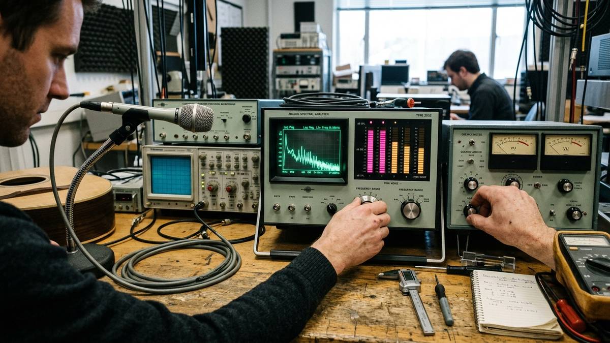
A spectrum is a recipe: it says where the energy sits across the audible range, from felt vibration to mosquito whine. White noise puts equal energy in every band — 1/f⁰, a flat line through the frequency domain. Pink noise declines three decibels each time the pitch interval doubles — 1/f¹. Brownian or red noise keeps falling, a steeper 6 dB per octave, 1/f², all thud and rumble. These are not flavors; they are slopes.
The ears, however, do not listen linearly, and the mismatch between exponent and periphery has consequences: hearing is calibrated so that equal-fold changes in frequency feel equal — the octave logic of logarithms — and its sensitivity drops steeply at extremes, so a flat spectrum concentrates perceived emphasis in the upper midrange where sibilants live. That is the famous hiss: white noise favors the sharp, rough textures the upper midrange contributes, because the ear’s amplification curve leans into exactly where the flat line does not taper.
Pink noise’s 3 dB/octave tilt intersects that sensitivity curve and cancels it almost perfectly: per octave the perceived level comes out roughly equal, which is why pink noise sounds like steady rain or a fan’s throb — and why a 1/f shape appears ubiquitously in nature: electroencephalography traces, heartbeats, music intervals follow that power law across timescales. This slope is the signature of long-range temporal correlations — statistics identical at every timescale — and natural signals, including the brain’s own rhythms, incline toward the pink: the physical preface to this acoustic comparison.
This 1/f universality runs deep enough to shape perception itself, which is why sounds seem louder in the dark and why a forest sounds restful while a data center does not: baseline expectations are matched by the tilted spectra the ears evolved inside, and mismatches register as effort. It also explains the nighttime asymmetry documented there — the brain lowers its auditory threshold after lights-out — so a masker tuned for daytime audition can misfire at night.
Acoustic Spectral Profile Comparison
- Core Focus:
- Neurochemistry:
- Dynamic:
Thalamic Sensory Gating: How Background Sound Silences the World
Sleep is a downgrade of consciousness and an upgrade of vulnerability, which is why the early brain evolved a security system. Along the thalamus — the sensory hub feeding the cortex — runs a sheet of inhibitory neurons called the thalamic reticular nucleus. Here the auditory gating question acquires its anatomical address. A footstep is a sudden, salient stimulus: the gate briefly fails, relay cells burst, the cortex receives the event, arousal chemistry is drafted. The white noise vs pink noise debate begins at the gate, before preference enters.
This gate is why the comparison must start in sleep neurophysiology rather than product reviews: the difference between the two spectra is not what each one says but what each one lets through. White noise vs pink noise is a question about the gate’s guest list.
Steady background sound attacks this problem three ways: it fills the dynamic range so only genuinely large events preserve contrast; it lowers the surprise statistic, since auditory neurons adapt to predictable input and stop transmitting it; and it caps the environment’s sensory contrast. Instead of many discrete noise events per hour — each a potential gate failure — there is one long, unvarying stimulus the machinery habituates to and stops transmitting: the masking mechanism behind acoustic color therapy.
Recommended Reading
Whether the masker should be flat or tilted is the pivot of the spectral noise question: one strategy covers ground, the other matches the sleeping brain’s own program.
But the evidence for masking is not uniformly rosy. In the ICU — one of the most sleep-destructive environments Medicine has built — steady white noise through headphones measurably reduced sleep disruptions and increased restful N2 [Stanchina et al., 2005], with a few patients sleeping worse on the noise nights. The stage masking protects — N2 — is the shallowest one, which raises the question the next section answers: can the right spectrum deepen rest rather than merely defend it?
The Northwestern Discovery: Pink Noise and Slow-Wave Enhancement
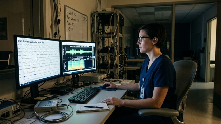
For decades, sleep-lab sound was defensive: reduce the noise, protect the rest. Then that reframed the field: the right acoustic stimulus could go on offense. At Northwestern University, Phyllis Zee’s laboratory demonstrated that timed pulses of pink noise, synced to the up-phase of the brain’s slow oscillations, amplified those oscillations and improved memory the next morning [Papalambros et al., 2017].
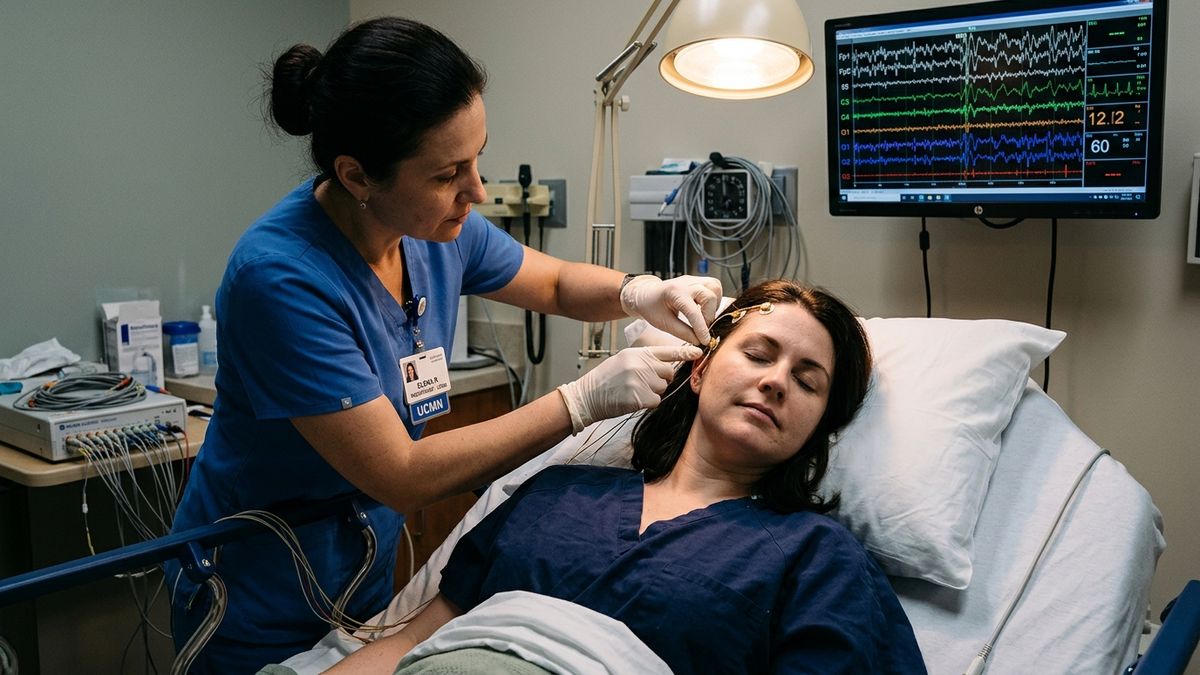
The mechanism is closed-loop phase-locked stimulation — the technical heart of this white noise vs pink noise distinction. An EEG sensor reads the brain’s slow rhythm in real time, and on each upswing a burst of pink noise is delivered, so the auditory thalamus perturbs the network precisely when the circuit is most excitable, and the slow rhythm strengthens instead of breaking.
The overnight picture was specific. Older adults recalled word pairs measurably better after a night of pink noise pulses, the improvement tracking each participant’s slow-wave amplification [Papalambros et al., 2017]. In younger adults, synchronizing pulses to the slow oscillation’s up-phase strengthened recall [Ngo et al., 2013], modeled by a 2012 companion study as a coupled oscillator pair — pink-noise source and EEG slow-wave generator — converging on a shared attractor [Zhou et al., 2012]. This electrophysiological entrainment forms the empirical foundation for closed-loop auditory sleep stimulation.
White Noise vs Pink Noise: The Neurological Trade-Off
When White Noise Wins: The Masking Case
The modern white noise vs pink noise comparison is a trade-off between two neuroscientific jobs. White noise is the blunt instrument of masking: if the bedroom’s problem is intermittent sound — traffic bursts, a snoring partner — the response is to fill the contrast gap, and a flat spectrum fills it from the lowest bass with equal uninterestingness across the range. In the ICU study, white noise protected N2 sleep against the worst acoustic territory Medicine has built.
For the high-arousal sleeper in the urban apartment or hospital ward, white noise’s constant, high-band hiss flattens the contrast a door slam would otherwise carry — the acoustic contrast divergence, made geographic.
When Pink Noise Wins: The Synchronization Case
Pink noise, by contrast, is the precise instrument. It masks weakly in the high band, but its genius is upstream, in entrainment: it sits where the brain’s own slow rhythms stand, and at low, non-arousing intensities the spectral performance gap widens — one supports rather than supplants them.
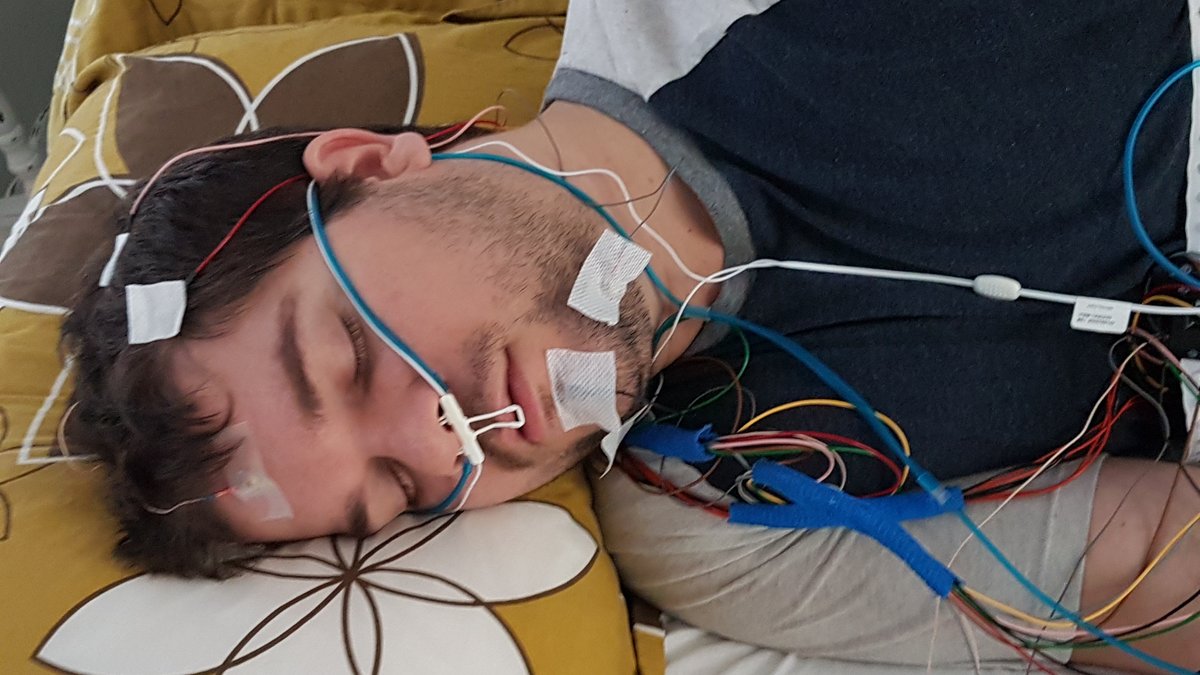
Clinical investigations in noise-heavy hospital environments (such as intensive care units) demonstrate that acoustic masking elevates arousal thresholds and stabilizes sleep continuity [Stanchina et al., 2005]. Furthermore, autonomic monitoring shows steady auditory masking reduces pulse rate variability triggered by sudden noise spikes. In older adults, white noise vs pink noise pulses — pink synchronized to slow oscillations — raised slow-wave power and improved word-pair recall over a sham night [Papalambros et al., 2017], and closed-loop acoustic stimulation during a single night significantly enhanced slow oscillations and boosted morning declarative memory recall in younger adults [Ngo et al., 2013]. Where white noise’s value ends at masking, white noise primarily functions as sensory masking against sudden environmental arousal, whereas pink noise research investigates active slow-wave electrophysiological modulation.
The white noise vs pink noise contrast differs not only in what the two spectra mask but in what they leave intact after the first slow wave of the night.
This tilt-match is a deep rule: biological systems, from DNA sequences to river networks to the resting heartbeat, exhibit 1/f fluctuations. The brain’s predictive machinery is tuned to that slope because it is the slope of nature — of music’s tempo fluctuations, of heartbeats, of the EEG itself — which is why the acoustic contrast is so visible in subjective reports: the flat spectrum reads as effort, the tilted one as environment. When the external spectrum matches the internal one, the brain’s resources shift from filtering noise to further synchronizing with it. Masking changes what reaches the gate; entrainment changes what the gate transmits next.
“All sleep sound machines are interchangeable as long as they block outside noise.”
White noise flattens acoustic contrast across high frequencies to suppress sudden awakenings, while pink noise (1/f) matches fractal brain dynamics to actively enhance delta slow-waves and overnight memory consolidation.
Decibels, Hair Cells, and the Habituation Ceiling: The Safe Usage Protocol
Every masking strategy has a ceiling, and the ceiling is the cochlea. Outer hair cells transduce sound into nerve impulses and do not regenerate: they have two states, functional and lost. Above roughly 85 dBA of sustained exposure, noise causes cumulative damage, and an 8-hour night is eight continuous hours of it. Consumer devices routinely exceed safe thresholds — analysis of infant sleep machines found maximum levels exceeding hospital-quiet limits — so the ceiling is decided at the pillow, not the spec sheet.
White noise vs pink noise changes not the risk class but its economics, and nightly use is a calculated risk. With chronic exposure the masking stimulus becomes part of the sleep-onset routine itself, and some habitual users cannot sleep without the device. Habituation decay compounds it: midbrain auditory neurons shift their responsiveness, the benefit erodes, users turn the volume up, walking into the cochlear dose zone.
Between them, a branch of the white noise vs pink noise question resolves on safety grounds: hair-cell risk is mostly a high-frequency problem — the 3–8 kHz octave where cochlear sensitivity peaks — The 1/f slope’s rolloff concentrates energy where cochlear sensitivity is flattest — why a safe pink soundscape can feel loud as white noise.
The 60-Decibel Rule and the Dependency Check
The protocol distills to numbers: a ceiling near 60 dB(A) — conversation volume at the ear — measured at the pillow, source across the room. The cochlea counts dose in energy-time, so distance and duration matter more than the label on the box. If you wake naturally, use a timer: the second half of the night has no masking job left, and every remaining hour is pure, unpaid cochlear exposure.
Track the dependency signal directly: if you cannot fall asleep without the machine, the tool has crossed from medicine into habit, and tapering is the exit, not raising it. Where the sleep-pressure deficit persists into daytime — the state examined in why you feel tired after 8 hours of sleep — the masking route has reached its boundary.
Bedtime sound strategy also interacts with the neurology of sleep onset: the approach to NREM is a delicate dissociative narrowing — the loss of wake-motor influence that causes the falling-jerk at the edge of sleep, whose extreme case is exploding head syndrome. A harsh stimulus during that handoff is maximally disruptive; the volume rule is the range in which the thalamus sees a steady, boring world rather than a threatening one, and the stage-2 sleep spindle can happen.
And when the alarm rings, the question is whether the protocol’s protected currency — sleep pressure a well-masked, delta-rich night manages — was preserved. That currency is the same one depleted in why you feel tired after 8 hours of sleep — the most honest outcome measure this acoustic sleep protocol offers.
The audit list: volume below 60 dB(A), device across the room, a timer if you wake naturally, and repair the environment when it is the problem — a snoring partner at 70 dB defeats any masker. Track whether you are waking less or merely sleeping with the sound: if you cannot fall asleep without it, taper rather than escalate — this decibel-safety protocol has a dependency check built in.
One boundary deserves honesty: no consumer device replicates the EEG-synchronized closed-loop of the laboratory systems. If daytime fatigue persists — the sleep-pressure deficit examined in why the body signals fatigue before collapse — the acoustic route has limits, and the acoustic sleep toolkit deserves an exit as much as an entrance.
Acoustic Spectrum & Neuro-Somnology Modality Matrix
The matrix below places the four acoustic environments side by side across the five parameters that decide sleep outcomes. The columns differ not in loudness but in architecture — white noise vs pink noise, read top to bottom, is the whole argument of this article in one frame.
| Spectral Environment | Flat White Noise (1/f⁰) | Pink Noise (1/f¹) | Brownian / Red Noise (1/f²) | Ambient Silence (No Sound) |
|---|---|---|---|---|
| Power Spectral Density Formula | S(f) ∝ 1/f⁰ — flat; equal energy per frequency bin | S(f) ∝ 1/f¹ — 3 dB/octave decline; equal energy per octave | S(f) ∝ 1/f² — 6 dB/octave decline; energy concentrated in low frequencies | S(f) → near-zero across bands; only residual room tone |
| Octave Energy Attenuation | 0 dB per octave — each doubling of frequency carries equal energy | −3 dB per octave — each octave holds as much power as the two below it combined | −6 dB per octave — each octave holds a quarter of the one below | Not applicable — no source spectrum to attenuate |
| Primary Thalamic Masking Mechanism | Broadband contrast flattening: uniform floor masks intermittent spikes across all bands, mostly in the higher-midrange where hearing favors it | Balanced masking plus slow-rhythm compatible input: accepted by auditory adaptation as quasi-natural; supports phase-locked entrainment | Low-band dominant masking: suppresses bass-range disturbances but leaves the high band uncovered | None — temporary threshold shift during silence makes spikes maximally salient |
| EEG Delta Wave Coherence Impact | Neutral-to-slightly-fragmenting: N2 protected but deep-stage architecture unaided | Positive: slow-oscillation phase-locked pulses increase slow-wave power and conjugate memory benefit | Near-neutral: too little high-frequency information to entrain, too much low energy to fully mask | Baseline: natural delta program preserved but vulnerable to every environmental spike |
| Subjective Somnological Tolerability | Hiss-like; fatiguing over hours; requires habituation period | Anecdoted as “steady rain”; most consistently rated pleasant and non-arousing | Rumbly; masks bass; can feel “heavy” or oppressive at volume | Rated ideal by low-noise-environment sleepers; intolerable for high-stimulus-contrast environments |
Read down the rows: white noise vs pink noise analysis in five parameters — a flat spectrum earns its badge in masking, the 1/f slope in synchronization, the brown exponent trades coverage for calm. Acoustic spectra, resolved by parameters.
Auditory Neuroscience Takeaway: Choosing the Right Sound Profile
Comparing white and pink noise, in one sentence: two different instruments pointed at two different neural problems. White noise masks the contrast that wakes the thalamic gate, and its flat spectrum earns its keep in cities, ICUs, and snoring bedrooms — the sensory masking split, rendered as geography. Pink noise both masks and synchronizes: its 1/f slope raises the gating floor with a sound the auditory system was already tuned to, and timed pulses amplify slow-wave sleep and memory.
The evidence leans pink: same masking floor, better sleep architecture, a friendlier sound, a safer cochlear profile. But the environment decides the tool: for a sudden-noise problem, white noise’s broadband flattening wins; for a sleep-quality problem, pink noise’s 1/f geometry is the instrument the delta oscillator respects.
The decibel rule stands in both cases: under 60, across the room — and if you cannot sleep without the sound, the question is no longer which noise but what the silence has been replaced by.
References
- Papalambros, N.A., Santostasi, G., Malkani, R., Braun, R., Zee, P.C. & Paller, K.A. (2017). Acoustic enhancement of sleep slow oscillations and concomitant memory improvement in older adults. Frontiers in Human Neuroscience, 11, 109. DOI: 10.3389/fnhum.2017.00109 · PMID: 28337134
- Zhou, J., Liu, D., Li, X., Ma, J., Zhang, J. & Fang, J. (2012). Pink noise: effect on complexity synchronization of brain activity and sleep consolidation. Journal of Theoretical Biology, 306, 68-72. DOI: 10.1016/j.jtbi.2012.04.006 · PMID: 22726808
- Kawada, T. & Suzuki, S. (1991). Sleep deepening effect of steady pink noise. Journal of Sound and Vibration, 151(3), 407-414. DOI: 10.1016/0022-460X(91)90537-T
- Stanchina, M.L., Abu-Hijleh, M., Chaudhry, B.K., Carlisle, C.C. & Millman, R.P. (2005). The influence of white noise on sleep in subjects exposed to ICU noise. Sleep Medicine, 6(5), 423-428. DOI: 10.1016/j.sleep.2004.12.004 · PMID: 16139772
- Steriade, M. (2006). Grouping of brain rhythms in corticothalamic systems. Neuroscience, 137(4), 1087-1106. DOI: 10.1016/j.neuroscience.2005.10.029 · PMID: 16343791
- Ngo, H.-V., Martinetz, T., Born, J. & Mölle, M. (2013). Auditory closed-loop stimulation of the sleep slow oscillation enhances memory. Neuron, 78(3), 545-553. DOI: 10.1016/j.neuron.2013.03.006 · PMID: 23583623
- Dang-Vu, T.T., McKinney, S.M., Buxton, O.M., Solet, J.M. & Ellenbogen, J.M. (2010). Spontaneous brain rhythms predict sleep stability in the face of noise. Current Biology, 20(15), R626-R627. DOI: 10.1016/j.cub.2010.06.032 · PMID: 20692606
📚 You Might Also Like
Hand-picked articles for you